濟(jì)南維修熱線
13153199508
長(zhǎng)沙維修熱線:13873135765
歡迎來(lái)到匠仁醫(yī)療設(shè)備有限公司網(wǎng)站!
濟(jì)南維修熱線
13153199508
長(zhǎng)沙維修熱線:13873135765

相信許多人都會(huì)有疑問(wèn),電子內(nèi)窺鏡貴,為什么還要做成一次性的?為什么能夠做成一次性的?讓我們一起來(lái)扒一扒它的出生,看一看它的經(jīng)歷。
Many people may wonder why electronic endoscopes, which are the most expensive, are still made disposable? Why can it be made disposable? Let's take a look at its birth and its experiences together.
在內(nèi)窺鏡200多年的發(fā)展歷程中,出現(xiàn)了幾次重大技術(shù)突破,將內(nèi)窺鏡帶入了不同的發(fā)展階段:整體經(jīng)歷了早期硬管式內(nèi)窺鏡、半可屈式內(nèi)窺鏡、纖維內(nèi)窺鏡、CCD電子鏡以及CMOS電子鏡四個(gè)階段。還有些人會(huì)把膠囊內(nèi)窺鏡列入第五階段,這是我們現(xiàn)階段正在經(jīng)歷的轉(zhuǎn)變,能不能列為第五階段還有待商榷。所以,在此我就把膠囊內(nèi)窺鏡列入創(chuàng)新發(fā)展里。同樣,許多其他新技術(shù)也列入該類進(jìn)行討論。
In the more than 200 years of development of endoscopes, there have been several major technological breakthroughs that have brought endoscopes into different stages of development: they have gone through four stages: early rigid tube endoscopes, semi flexible endoscopes, fiber endoscopes, CCD electronic microscopes, and CMOS electronic microscopes. Some people still classify capsule endoscopes as the fifth stage, which is a transformation we are currently experiencing. Whether it can be classified as the fifth stage remains to be discussed. So, I will include capsule endoscopy in the innovative development here. Similarly, many other new technologies are also included in this category for discussion.
01內(nèi)窺鏡的發(fā)展歷程
The Development History of 01 Endoscope
階段:光學(xué)硬鏡、半屈式內(nèi)鏡(1806年起) 1806年,德國(guó)醫(yī)生Philip Bozzini在病人的肛門內(nèi)插入一根硬管,借助于蠟燭的光亮,觀察膀胱和直腸內(nèi)部病變,他將其稱為"LICHTLEITER",這被認(rèn)為是內(nèi)窺鏡發(fā)展史的開端。但當(dāng)時(shí)的人們并不理解這種檢查方法, Bozzini 也因他的“好奇心”受到維也納 醫(yī)學(xué)院的處罰。次將"LICHTLEITER”運(yùn)用于人體的是法國(guó)外科醫(yī)生 Desormeaux因此他被許多人譽(yù)為“內(nèi)窺鏡之父”。他的“LICHTLEITER”是以燒煤油和松節(jié)油的燈為光源,燈的上方帶有煙囪并用透鏡將光線聚集以增加亮度,可想而知灼傷是進(jìn)行這種檢查時(shí)的主要并發(fā)癥。雖然這種內(nèi)窺鏡可以到達(dá)胃,但光線太暗,所以“LICHTLEITER”主要用于檢查泌尿系方面的疾病。1868 年,Desormeaux 和 Segelar 次在一篇文章中使用“內(nèi)窺鏡”一詞。Philip Bozzini的“LICHTLEITER”
In 1806, German doctor Philip Bozzini inserted a hard tube into the patient's anus and used the light of a candle to observe the internal lesions of the bladder and rectum. He called it the "LICHTLEITER", which is considered the beginning of the history of endoscopic development. But at that time, people did not understand this examination method, and Bozzini was also punished by the Vienna Medical School for his "curiosity". The first time the "LICHTLEITER" was applied to the human body was by French surgeon Desormeaux, who is widely known as the "father of endoscopes". His "LICHTLEITER" uses a kerosene and turpentine burning lamp as a light source, with a chimney above the lamp and a lens to concentrate the light to increase brightness. It can be imagined that burns are the main complication of this examination. Although this endoscope can reach the stomach, the light is too dim, so "LICHTLEITER" is mainly used to examine urinary system diseases. In 1868, Desormeaux and Segelar first used the term "endoscope" in an article. Philip Bozzini's' LICHTLEITER '
1932年,德國(guó)人Schindler與Wolf成功研制出一種半屈式內(nèi)鏡,定名為 Wolf-Schindler式胃鏡。這種內(nèi)鏡由48個(gè)透鏡組成光學(xué)系統(tǒng),燈泡光亮度強(qiáng),醫(yī)生能清晰地觀察圖像。真正意義上的個(gè)半可屈式胃窺鏡是由 Schindler從 1928 年起與的器械制作師 Wolf 合作開始研制的 并終在1932年獲得成功 該胃鏡直徑為12mm長(zhǎng)為 77cm,光學(xué)系統(tǒng)由 48 個(gè)透鏡組成,前端部分可在不同水平面彎曲34°,即在胃內(nèi)有一定范圍的彎曲,使術(shù)者能清晰地觀察胃粘膜圖像,該胃鏡前端有一光滑金屬球,插入較方便,燈泡光亮度較強(qiáng),有空氣通道用以注氣,近端為硬管部,有接目鏡調(diào)焦。
In 1932, German Schindler and Wolf successfully developed a half flexion endoscope, which was named Wolf Schindler gastroscope. This endoscope consists of an optical system consisting of 48 lenses, and the light bulb has strong brightness, allowing doctors to observe images clearly. The first semi flexible gastroscope in the true sense was developed by Schindler in collaboration with the excellent instrument maker Wolf since 1928, and finally succeeded in 1932. The diameter of the gastroscope is 12mm and the length is 77cm. The optical system consists of 48 lenses, and the front end can bend 34 ° at different horizontal planes, which means there is a certain range of curvature in the stomach, allowing the surgeon to clearly observe the image of the gastric mucosa. The front end of the gastroscope has a smooth metal ball, which is easy to insert, and the light bulb has strong brightness. There is an air channel for gas injection, and the proximal end is a hard tube part with a focusing eyepiece.Wolf-Schindler 式胃鏡的創(chuàng)制,開辟了胃鏡檢查術(shù)的新紀(jì)元。
Schindler與Wolf的Wolf-Schindler式胃鏡
Schindler and Wolf's Wolf Schindler style gastroscopy
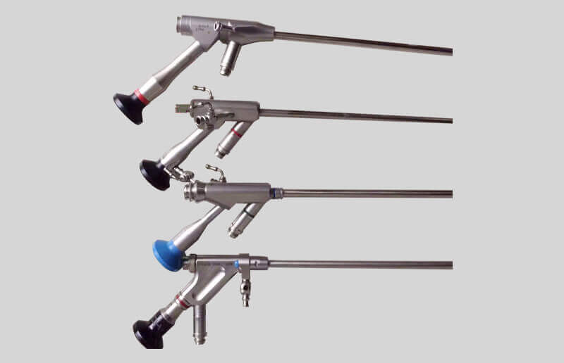
1960年,卡爾史托斯公司成功研發(fā)冷光源;1965年,卡爾史托斯公司研發(fā)推廣HOPKINS柱狀透鏡技術(shù),直今天,硬性光學(xué)鏡仍使用這一結(jié)構(gòu)。
In 1960, Karl Storz successfully developed a cold light source; In 1965, Karl Storz developed and promoted the HOPKINS cylindrical lens technology, which is still used in rigid optical mirrors today.
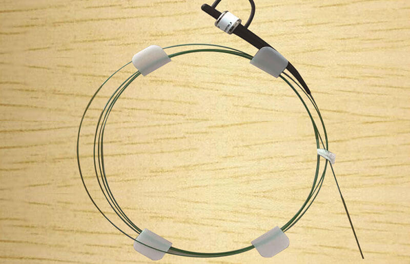
第二階段:光纖內(nèi)鏡(1957年起) 1954年,基于之前纖維傳導(dǎo)光源及纖維間加蓋覆層可以纖維間光絕緣的理論基礎(chǔ),英國(guó)Hopkings及Kapany研究了纖維的精密排列,有效地了纖維束的圖像傳遞,為纖維光學(xué)的實(shí)用奠定了基礎(chǔ)。內(nèi)窺鏡實(shí)現(xiàn)了一大歷史性突破,應(yīng)用光纖傳像的內(nèi)鏡登上舞臺(tái)。 1957年,Curtiss和Hirschowitz把大量的玻璃纖維薪合成一束,并在末端把這些纖維融合在一起,以便使它們根據(jù)自身的長(zhǎng)度獨(dú)立活動(dòng),就此造就了個(gè)軟質(zhì)內(nèi)鏡。Hirschowitz和他的研究組制成了世界上個(gè)用于檢查胃、十二指腸的光導(dǎo)纖維內(nèi)鏡原型。 1960年,美國(guó)膀胱鏡制造者公司(ACMI,現(xiàn)在年輕人應(yīng)該都不知道這個(gè)了,都成傳說(shuō)了)向Hirschowitz提供了個(gè)商業(yè)纖維內(nèi)窺鏡。1964年,Olympus 廠在光導(dǎo)纖維胃鏡基礎(chǔ)上,加裝了活裝置及照相機(jī),有效地顯示了胃照相術(shù),實(shí)時(shí)觀察胃內(nèi)狀況。1966 年 Olympus 廠首創(chuàng)前端彎角機(jī)構(gòu)。1967年,Machida廠采用外部冷光源,使光亮度大增,可發(fā)現(xiàn)小病灶,視野進(jìn)一步擴(kuò)大,可以觀察到十二指腸。隨著附屬裝置的不斷改進(jìn),如手術(shù)器械、攝影系統(tǒng)的發(fā)展,內(nèi)鏡不但可用于診斷,且可用于手術(shù),這意味著微創(chuàng)手術(shù)時(shí)代的到來(lái)。
Phase 2: Fiber Optic Endoscopy (since 1957) In 1954, based on the theoretical foundation that fiber conductive light sources and fiber to fiber cladding could solve fiber to fiber optical insulation, Hopkins and Kapany from the UK studied the precise arrangement of fibers, effectively solving the image transmission of fiber bundles and laying the foundation for the practical application of fiber optics. Endoscopy has achieved a historic breakthrough, with endoscopes utilizing fiber optic imaging taking the stage. In 1957, Curtiss and Hirschowitz combined a large amount of glass fibers into a bundle and fused these fibers together at the end to allow them to move independently according to their own length, thus creating the first soft endoscope. Hirschowitz and his research team have developed the world's first prototype of fiber optic endoscopy for examining the stomach and duodenum. In 1960, the American cystoscope manufacturer company (ACMI, now a legendary brand unknown to young people) provided Hirschowitz with the first commercial fiber endoscope. In 1964, Olympus factory added live devices and cameras on the basis of fiber optic gastroscopy, effectively displaying gastric photography and observing the condition of the stomach in real time. In 1966, Olympus factory pioneered the front-end bending mechanism. In 1967, Machida factory adopted an external cold light source, which greatly increased the brightness and allowed for the detection of small lesions, further expanding the field of view and allowing observation of the duodenum. With the continuous improvement of accessory devices such as surgical instruments and photography systems, endoscopes can not only be used for diagnosis, but also for surgical treatment, which means the arrival of the era of minimally invasive surgery.
Hirschowitz醫(yī)生
Dr. Hirschowitz
第三階段:CCD電子內(nèi)鏡(1983年起) 1983 年美國(guó) Welch Allyn 公司研制并應(yīng)用微型圖像傳感器(charge coupled device,CCD)代替了內(nèi)鏡的光導(dǎo)纖維導(dǎo)像術(shù),宣告了電子內(nèi)鏡的誕生--內(nèi)鏡發(fā)展史上另一次歷史性的突破。電子內(nèi)窺鏡主要由內(nèi)鏡(endoscopy)、電視信息系統(tǒng)中心(video information system center)和電視監(jiān)視器(television monitor)三個(gè)主要部分組成,另外還配備一些輔助裝置,如錄像機(jī)、照相機(jī)、吸引器以及用來(lái)輸入各種信息的鍵盤和診斷所用的各種處置器具等。它的成像主要依賴于鏡身前端裝備的 CCD,CCD 就像一臺(tái)微型攝像機(jī)將圖像經(jīng)過(guò)圖像處理器處理后,顯示在電視監(jiān)視器的屏幕上。比普通光導(dǎo)纖維內(nèi)鏡的圖像清晰,色澤逼真,分辨率更高,而且可供多人同時(shí)觀看。由于電子內(nèi)鏡的問(wèn)世,給百余年來(lái)內(nèi)鏡的診斷和開創(chuàng)了歷史新篇章,在臨床、教學(xué)和科研中發(fā)揮出它巨大的優(yōu)勢(shì)。隨后,日本的富士、奧林巴斯(1985年推出其電子內(nèi)窺鏡系統(tǒng) EVIS-1)等企業(yè)相繼宣告電子內(nèi)鏡的成功研制。從像素?cái)?shù)量上看,光纖內(nèi)鏡的分辨率一般為2萬(wàn)像素,而應(yīng)用CCD的電子鏡分辨率一般是光纖內(nèi)鏡的20倍,這使得電子內(nèi)鏡的圖像質(zhì)量、細(xì)節(jié)信息、清晰程度都有了極大提升,基于內(nèi)窺鏡的診療技術(shù)開啟了新的篇章。上世紀(jì)70~90年代,全球超過(guò)半數(shù)的半導(dǎo)體在日本生產(chǎn),全球前10大半導(dǎo)體公司中,日本一度獨(dú)占6家,這使得日本的內(nèi)鏡企業(yè)發(fā)展優(yōu)勢(shì)。
Phase Three: CCD Electronic Endoscopy (Since 1983) In 1983, Welch Allyn Company in the United States developed and applied a miniature image sensor (charge coupled device, CCD) to replace the optical fiber guidance of endoscopy, announcing the birth of electronic endoscopy - another historic breakthrough in the history of endoscopy development. Electronic endoscopes are mainly composed of three main parts: endoscopy, video information system center, and television monitor. In addition, they are equipped with auxiliary devices such as video recorders, cameras, suction devices, keyboards for inputting various information, and various treatment equipment used for diagnosis and treatment. Its imaging mainly relies on the CCD equipped at the front end of the mirror body. The CCD is like a miniature camera that processes the image through an image processor and displays it on the screen of a television monitor. Compared to ordinary fiber optic endoscopes, the image is clearer, the color is realistic, the resolution is higher, and it can be viewed by multiple people at the same time. Due to the emergence of electronic endoscopy, a new chapter in the history of endoscopic diagnosis and treatment has been opened for more than a hundred years, and it has played a huge advantage in clinical, teaching, and scientific research. Subsequently, Japanese companies such as Fuji and Olympus (which launched their first electronic endoscope system EVIS-1 in 1985) successively announced the successful development of electronic endoscopes. In terms of pixel count, the resolution of fiber optic endoscopes is generally 20000 pixels, while the resolution of electronic endoscopes using CCD is generally 20 times that of fiber optic endoscopes. This greatly improves the image quality, detail information, and clarity of electronic endoscopes, opening a new chapter in diagnosis and treatment technology based on endoscopes. In the 1970s and 1990s, more than half of the world's semiconductors were produced in Japan. Among the top 10 semiconductor companies in the world, Japan once monopolized 6, which gave Japanese endoscope companies a great development advantage.
CCD
CCD
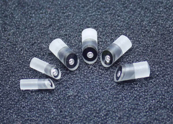
第四階段:CMOS電子內(nèi)鏡(1995年起) 1995年,幾位華人留學(xué)生在硅谷創(chuàng)立豪威,這家企業(yè)實(shí)現(xiàn)了此后CMOS(Complementary Metal Oxide Semiconductor,互補(bǔ)金屬氧化物半導(dǎo)體,一種集成電路的設(shè)計(jì)工藝)技術(shù)對(duì)圖像傳感器市場(chǎng)的破局。在當(dāng)時(shí)的工藝條件下,CMOS的成像質(zhì)量相較CCD稍差,但卻可以做到功耗和成本的極大降低。隨著索尼、三星等傳統(tǒng)半導(dǎo)體巨頭的加入和CMOS生產(chǎn)工藝的精進(jìn),CMOS圖像傳感器的性能逐漸提高,在內(nèi)窺鏡中的應(yīng)用研究也逐漸展開。與CCD相比,CMOS傳感器具有成本低、集成度高、耗電量小等優(yōu)點(diǎn)。 2015年5月美國(guó)豪威被由中信資本、北京清芯華創(chuàng)和金石投資組成的財(cái)團(tuán)以19億美元收購(gòu),終于2016年初完成私有化,成為北京豪威的全資子公
Phase Four: CMOS Electronic Endoscope (Since 1995) In 1995, several Chinese international students founded Howay in Silicon Valley, which achieved the breakthrough of CMOS (Complementary Metal Oxide Semiconductor, a design process for integrated circuits) technology in the image sensor market. Under the process conditions at that time, the imaging quality of CMOS was slightly inferior to CCD, but it could achieve significant reductions in power consumption and cost. With the addition of traditional semiconductor giants such as Sony and Samsung and the improvement of CMOS production processes, the performance of CMOS image sensors is gradually improving, and research on the application of endoscopes is also gradually unfolding. Compared with CCD, CMOS sensors have the advantages of low cost, high integration, and low power consumption. In May 2015, American Howe was acquired by a consortium consisting of CITIC Capital, Beijing Qingxin Huachuang, and Jinshi Investment for $1.9 billion. It was eventually privatized in early 2016 and became a wholly-owned subsidiary of Beijing Howe
CMOS
CMOS
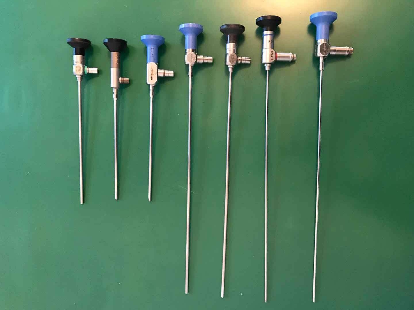
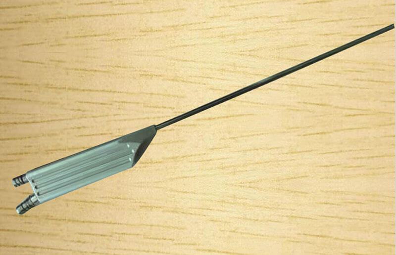
目前,市場(chǎng)上的軟鏡主要使用電子鏡方案,CMOS攝像頭在鏡體前端,鏡體可彎曲,鏡體后端連接圖像處理裝置;硬鏡中電子鏡與光學(xué)鏡兩種方案并存,電子硬鏡的優(yōu)勢(shì)是不像光學(xué)鏡易損壞、不需手動(dòng)調(diào)焦,而光學(xué)硬鏡的優(yōu)勢(shì)是透鏡組傳像的保真度高,CMOS安裝在鏡體后端不受鏡體直徑限制。
At present, the soft mirrors on the market mainly use electronic mirror solutions, with CMOS cameras at the front end of the mirror body, which can be bent, and image processing devices connected to the back end of the mirror body; There are two solutions for hard mirrors: electronic mirrors and optical mirrors. The advantage of electronic hard mirrors is that they are not as easily damaged and do not require manual focusing as optical mirrors, while the advantage of optical hard mirrors is that the lens group has high fidelity in image transmission. CMOS is installed at the rear end of the mirror body and is not limited by the diameter of the mirror body.
總結(jié)
summary
CCD(電荷耦合器件)和CMOS(互補(bǔ)金屬氧化物半導(dǎo)體)是兩種不同類型的圖像傳感器,它們各有特點(diǎn)和優(yōu)勢(shì),以下是CCD和CMOS的主要區(qū)別及各自的優(yōu)點(diǎn):CCD(Charge-Coupled Device)工作原理:CCD通過(guò)一個(gè)像素傳遞到另一個(gè)像素的方式來(lái)收集光,并將其轉(zhuǎn)換成電子信號(hào)。圖像質(zhì)量:通常提供高質(zhì)量的圖像,具有的光學(xué)特性和較低的噪點(diǎn)。靈敏度:對(duì)光線的響應(yīng)較好,適合低光照條件下拍攝。能耗:相對(duì)較高,因?yàn)樗邢袼囟际峭ㄟ^(guò)一個(gè)輸出節(jié)點(diǎn)來(lái)讀取。CMOS(Complementary Metal-Oxide-Semiconductor)工作原理:CMOS傳感器為每個(gè)像素配備了一個(gè)放大器,可以在像素處直接轉(zhuǎn)換光信號(hào)為電信號(hào)。成本和制造:制造成本較低,因?yàn)樗梢允褂脴?biāo)準(zhǔn)的半導(dǎo)體制造工藝。能耗:相比CCD,CMOS的能耗較低,更適合用于便攜設(shè)備如智能手機(jī)。速度:數(shù)據(jù)讀取速度更快,適合高速攝影和視頻錄制。CCD的優(yōu)點(diǎn):提供高質(zhì)量的圖像,特別是在色彩重現(xiàn)、動(dòng)態(tài)范圍和低光照條件下的性能方面。CMOS的優(yōu)點(diǎn):成本較低,能耗低,數(shù)據(jù)處理速度快,更適合快速拍攝和長(zhǎng)時(shí)間使用。
CCD (Charge Coupled Device) and CMOS (Complementary Metal Oxide Semiconductor) are two different types of image sensors, each with its own characteristics and advantages. The main differences and advantages of CCD and CMOS are as follows: CCD (Charge Coupled Device) works by collecting light from one pixel to another and converting it into electronic signals. Image quality: Typically provides high-quality images with excellent optical properties and low noise. Sensitivity: Good response to light, suitable for shooting under low light conditions. Energy consumption: relatively high because all pixels are read through one output node. The working principle of CMOS (Complementary Metal Oxide Semiconductor): CMOS sensors are equipped with an amplifier for each pixel, which can directly convert optical signals into electrical signals at the pixel. Cost and Manufacturing: The manufacturing cost is lower because it can use standard semiconductor manufacturing processes. Energy consumption: Compared to CCD, CMOS has lower energy consumption and is more suitable for portable devices such as smartphones. Speed: Faster data reading speed, suitable for high-speed photography and video recording. Advantages of CCD: Provides high-quality images, especially in terms of color reproduction, dynamic range, and performance under low light conditions. Advantages of CMOS: Low cost, low energy consumption, fast data processing speed, more suitable for fast shooting and long-term use.
隨著技術(shù)的發(fā)展,CMOS傳感器的性能已經(jīng)大幅提升,逐漸在許多應(yīng)用領(lǐng)域取代了CCD傳感器,特別是在消費(fèi)級(jí)數(shù)碼相機(jī)和手機(jī)攝像頭中。然而,CCD仍然在一些和科學(xué)應(yīng)用中保持著其優(yōu)勢(shì)地位。選擇哪種類型的傳感器取決于具體的應(yīng)用需求、預(yù)算和期望的圖像質(zhì)量。
With the development of technology, the performance of CMOS sensors has been greatly improved, gradually replacing CCD sensors in many application fields, especially in consumer digital cameras and mobile phone cameras. However, CCD still maintains its advantageous position in some professional and scientific applications. The choice of which type of sensor depends on specific application requirements, budget, and expected image quality.
顯然,一開始大家所熟知的電子內(nèi)窺鏡昂貴主要是由于CCD昂貴,CCD質(zhì)量越好,呈現(xiàn)畫面質(zhì)量越高,也越高。后來(lái)CMOS出現(xiàn)了,成本比CCD還低,且于16年豪威完成私有化,大幅降低了國(guó)內(nèi)企業(yè)的采購(gòu)成本。這就解釋了為什么一次性內(nèi)窺鏡采用的是電子芯片,而且也是近幾年一次性內(nèi)窺鏡在國(guó)內(nèi)蓬勃發(fā)展的主要原因。
Obviously, the well-known high cost of electronic endoscopes at the beginning was mainly due to the high cost of CCD. The better the quality of CCD, the higher the image quality presented, and the higher the price. Later on, CMOS emerged with lower costs than CCD, and in 2016, Huawei completed privatization, significantly reducing the procurement costs of domestic enterprises. This explains why disposable endoscopes use electronic chips, and it is also the main reason for the booming development of disposable endoscopes in China in recent years.
本文由 內(nèi)窺鏡設(shè)備維修 友情奉獻(xiàn).更多有關(guān)的知識(shí)請(qǐng)點(diǎn)擊 http://www.dcgr.cn/ 真誠(chéng)的態(tài)度.為您提供為的服務(wù).更多有關(guān)的知識(shí)我們將會(huì)陸續(xù)向大家奉獻(xiàn).敬請(qǐng)期待.
This article is a friendly contribution from the maintenance of endoscopic equipment. For more related knowledge, please click http://www.dcgr.cn/ Sincere attitude. We provide you with comprehensive services. We will gradually contribute more relevant knowledge to everyone. Please stay tuned
 新聞資訊
新聞資訊 聯(lián)系我們
 公司:匠仁醫(yī)療設(shè)備有限公司
  新聞資訊
新聞資訊微信公眾號(hào)

樊經(jīng)理:13153199508 李經(jīng)理:13873135765
公司地址:山東省濟(jì)南市槐蔭區(qū)美里東路3000號(hào)德邁國(guó)際信息產(chǎn)業(yè)園6號(hào)樓101-2室 湖南省長(zhǎng)沙市雨花區(qū)勞動(dòng)?xùn)|路820號(hào)恒大綠洲14棟2409室
魯公網(wǎng)安備 37010402441281號(hào)